Neurobiology of Resilience
In the quest to understand resilience – the psychological fortress that empowers individuals to rebound and even flourish amid adversity–we delve deep into the realm of the human brain, the epicenter of stress and resilience. This chapter illuminates the intricate tapestry of neurobiological processes underscoring resilience, demystifying the dance between stress, brain structures, and mechanisms fostering resilience.
Brain Structures and Their Functions
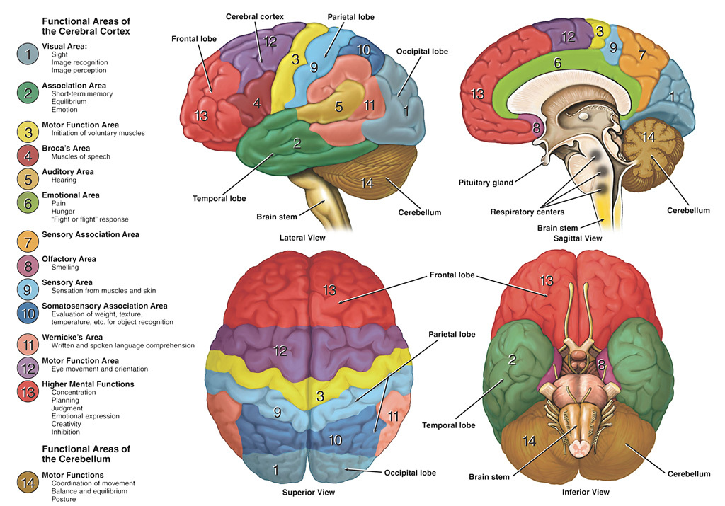
The human brain is an intricate organ composed of billions of neurons and glial cells, responsible for the complexity of human thought, emotions, and behaviors. The various structures of the brain work together in complex intricate manners to process information, regulate bodily functions, and interact with the environment.
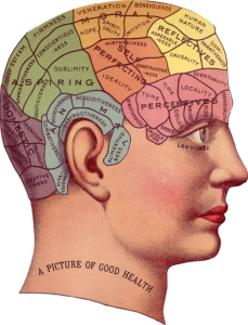
Major Structures and Their Functions
The Cerebrum
The cerebrum is the largest part of the brain and is divided into two hemispheres (left and right). It is responsible for integrating sensory information, initiating motor functions, and facilitating complex cognitive processes such as learning, memory, and reasoning (Bear, Connors, & Paradiso, 2020).
Lobes of the Cerebrum
- Frontal Lobe: Involved in decision making, problem solving, and planning. It houses the primary motor cortex, which controls voluntary movements.
- Parietal Lobe: Processes sensory information from the body, including touch, temperature, and pain. It also plays a role in spatial orientation and manipulation.
- Temporal Lobe: Essential for auditory processing and is also involved in memory and emotion through the hippocampus and amygdala.
- Occipital Lobe: Dedicated to vision, interpreting information from the eyes.
The Cerebellum
Located under the cerebrum, the cerebellum is critical for motor control, coordination, precision, and accurate timing of movements. It also plays a role in cognitive functions such as attention and language (Schmahmann, 2019).
The Brainstem
The brainstem connects the cerebrum and cerebellum to the spinal cord and is made up of the midbrain, pons, and medulla oblongata. It regulates vital functions such as heart rate, breathing, and sleep cycles (Augustine et al., 2023).
The Limbic System
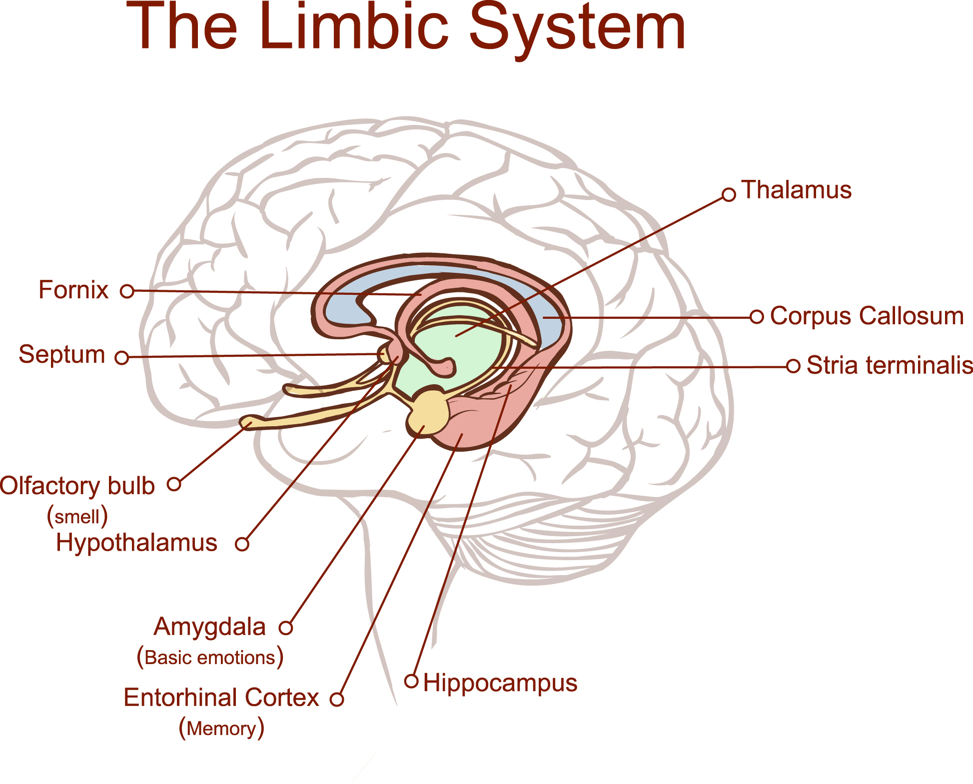
The limbic system is a complex system of nerves and networks in the brain, involving several areas near the edge of the cortex concerned with instinct and mood. It controls basic emotions (fear, pleasure, anger) and drives (hunger, sex, dominance, care of offspring) (LeDoux, 2000).
- Hippocampus: Essential for learning and memory formation.
- Amygdala: Plays a crucial role in emotion processing and response.
- Thalamus: Acts as the brain’s relay station, directing sensory and motor signals to the cerebral cortex.
- Hypothalamus: Regulates homeostasis, including temperature, hunger, thirst, and circadian rhythms.
Limbic System and Resilience
The limbic system is a complex network of structures in the brain that plays an essential role in regulating emotions, forming memories, and governing behaviors. At the heart of this system are the amygdala and the hippocampus, two almond-shaped structures that have garnered significant attention in neuroscience for their pivotal roles in emotional regulation, memory, and resilience. A balance between the reactivity of the amygdala and the modulatory influence of the hippocampus is crucial for resilience, enabling individuals to respond to stressors without getting overwhelmed (Davidson & McEwen, 2012).
The amygdala, often dubbed the ’emotional sentinel’ of the brain, primarily functions in detecting, processing, and responding to emotionally salient stimuli, especially those that are perceived as threatening or rewarding (LeDoux, 2000). When confronted with a potential threat, the amygdala quickly evaluates the situation and determines the appropriate emotional response, be it fear, anger, or something else. It is also deeply intertwined with the body’s stress response system, which means that its activity can trigger physiological responses like increased heart rate, heightened alertness, or a rush of adrenaline. Over time, if the amygdala’s reactivity remains unchecked, it can predispose an individual to conditions like anxiety disorders or post-traumatic stress disorder (PTSD) where the brain becomes hyper-responsive to perceived threats (Shin & Liberzon, 2010).
On the other hand, the hippocampus is intricately involved in the formation and retrieval of memories, particularly those related to personal experiences and contexts, known as episodic memories (Eichenbaum, 2000). This structure enables the brain to contextualize events, allowing individuals to understand their experiences within a broader framework of past events. Furthermore, the hippocampus plays a modulating role in stress response. Research has indicated that chronic stress can lead to reduced hippocampal volume and subsequently impair its functioning. This impairment can lead to difficulties in distinguishing between safe and threatening contexts, which can exacerbate the heightened reactivity of the amygdala (Sapolsky, 2000).
For resilience, the balance between the amygdala’s reactivity and the hippocampus’s modulatory influence is vital, allowing an individual to cope with and adapt to stressors without becoming overwhelmed or maladaptive. When the amygdala and hippocampus function in harmony, they enable the brain to appropriately respond to stressors, integrating emotional reactions with past experiences, and facilitating adaptive responses. If this balance is skewed, either due to chronic stress, trauma, or other factors, it can result in increased vulnerability to psychological disorders and reduced resilience (Davidson & McEwen, 2012). As such, the interconnected roles of the amygdala and hippocampus within the limbic system underscore their importance in emotional regulation, memory formation, and resilience. A deeper understanding of these structures can provide valuable insights into the neurobiological underpinnings of human emotions and behaviors, and pave the way for novel therapeutic interventions for conditions rooted in limbic system dysfunctions.
Neuron Anatomy and Function
Neuron Anatomy
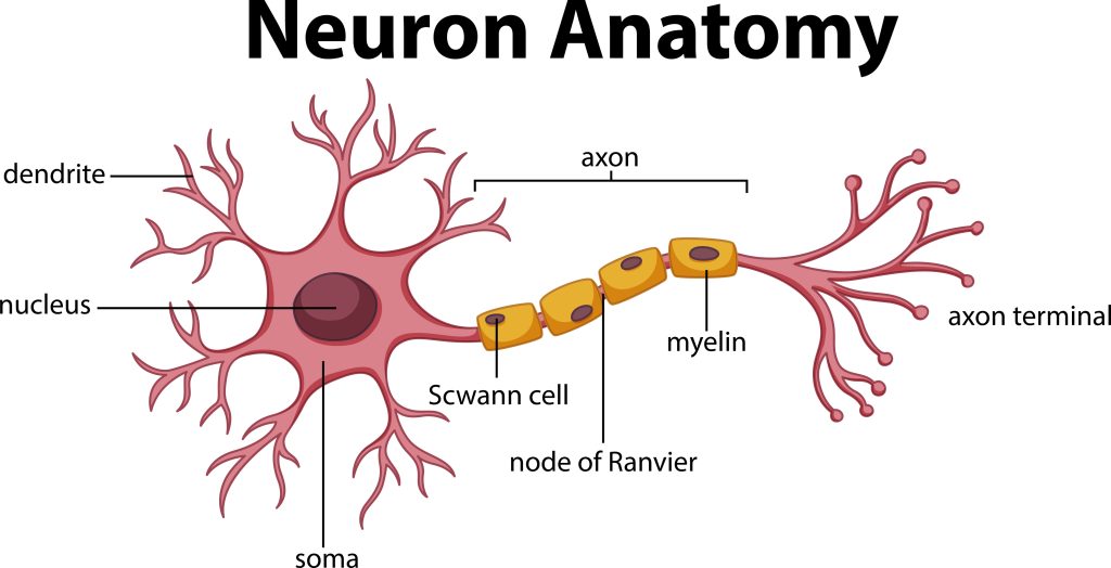
Understanding neuron anatomy is crucial for appreciating how the brain and the wider nervous system function. The specific architectural features of neurons facilitate the rapid and controlled transmission of information, ensuring that the body can respond appropriately to internal and external stimuli. The neuron, often referred to as a nerve cell, is the fundamental unit of the brain and nervous system, responsible for transmitting information throughout the body. A neuron’s structure is uniquely suited to its function, featuring several distinct parts that facilitate the transmission of electrical signals (Bear, Connors, & Paradiso, 2020). The main parts of a neuron include the cell body (soma), dendrites, axon, and synaptic terminals. The cell body houses the nucleus and cytoplasm, providing essential metabolic support and synthesizing proteins crucial for neuron function. Dendrites, branching off from the cell body, receive incoming signals from other neurons and conduct these signals towards the cell body. This input is then processed and, if sufficient, triggers the neuron to generate an electrical impulse.
The axon is a long, slender projection that extends from the cell body and transmits the electrical signal away from the cell body towards other neurons or muscles. Axons can vary dramatically in length, with some extending a few micrometers and others up to a meter or more in humans (Kandel, Schwartz, Jessell, Siegelbaum, & Hudspeth, 2013). Axons are often surrounded by a myelin sheath, a layer of fatty material composed of glial cells that insulates the axon, allowing electrical signals to travel faster and more efficiently. This myelination is critical for rapid signal transmission and affects the speed of reflexes and sensory processing. At the end of the axon are the synaptic terminals, which are specialized to release neurotransmitters, the chemical messengers that propagate the signal to the next neuron in the chain.
The interaction between neurons occurs at synapses, which are the gaps between the synaptic terminals of one neuron and the dendrites of another. When an electrical impulse reaches a synaptic terminal, it triggers the release of neurotransmitters into the synaptic cleft. These neurotransmitters then bind to receptors on the postsynaptic neuron, causing changes that can either excite or inhibit the neuron, depending on the type of neurotransmitter and receptor involved (Purves et al., 2018). This complex interaction allows the nervous system to carry out myriad functions, from simple reflexes to complex cognitive processes such as thinking and planning.
Neuron Anatomy and Resilience
The anatomy of neurons and their intricate connections are fundamentally related to the concept of neural resilience, which refers to the nervous system’s ability to adapt and recover from injuries, stress, or diseases. Understanding neuron anatomy provides insight into the mechanisms that underlie this resilience.
Neural Plasticity: One of the key aspects of resilience in the neural context is plasticity, which is the ability of neurons to change their connections and behavior in response to new information, sensory stimulation, development, damage, or dysfunction (Purves et al., 2018). For instance, after a traumatic brain injury, surrounding neurons can alter their pathways and create new synapses to compensate for lost functions. The dendrites and synaptic terminals are critical in these adaptive changes, as they are the main sites for synaptic modification and new connection formation.
Myelination: The myelin sheath that surrounds many axons plays a crucial role in the speedy and efficient transmission of electrical signals across long distances in the nervous system. After injury, the process of remyelination, or the reconstruction of the myelin sheath, is a pivotal aspect of neural resilience and recovery. Effective remyelination helps restore rapid communication between neurons, which is crucial for regaining functions that might have been impaired by the injury (Kandel et al., 2013).
Neurotransmitter Dynamics: The resilience of the neural system is also evident in the synaptic terminals’ ability to modulate neurotransmitter release in response to changes in the neural environment. After a neuron is damaged, the chemical balance at synapses can be disrupted. The capacity of neurons to adjust the release of neurotransmitters and modify receptor activity can help stabilize neural circuits, aiding recovery and maintaining functionality (Bear, Connors, & Paradiso, 2020).
Neurotransmission
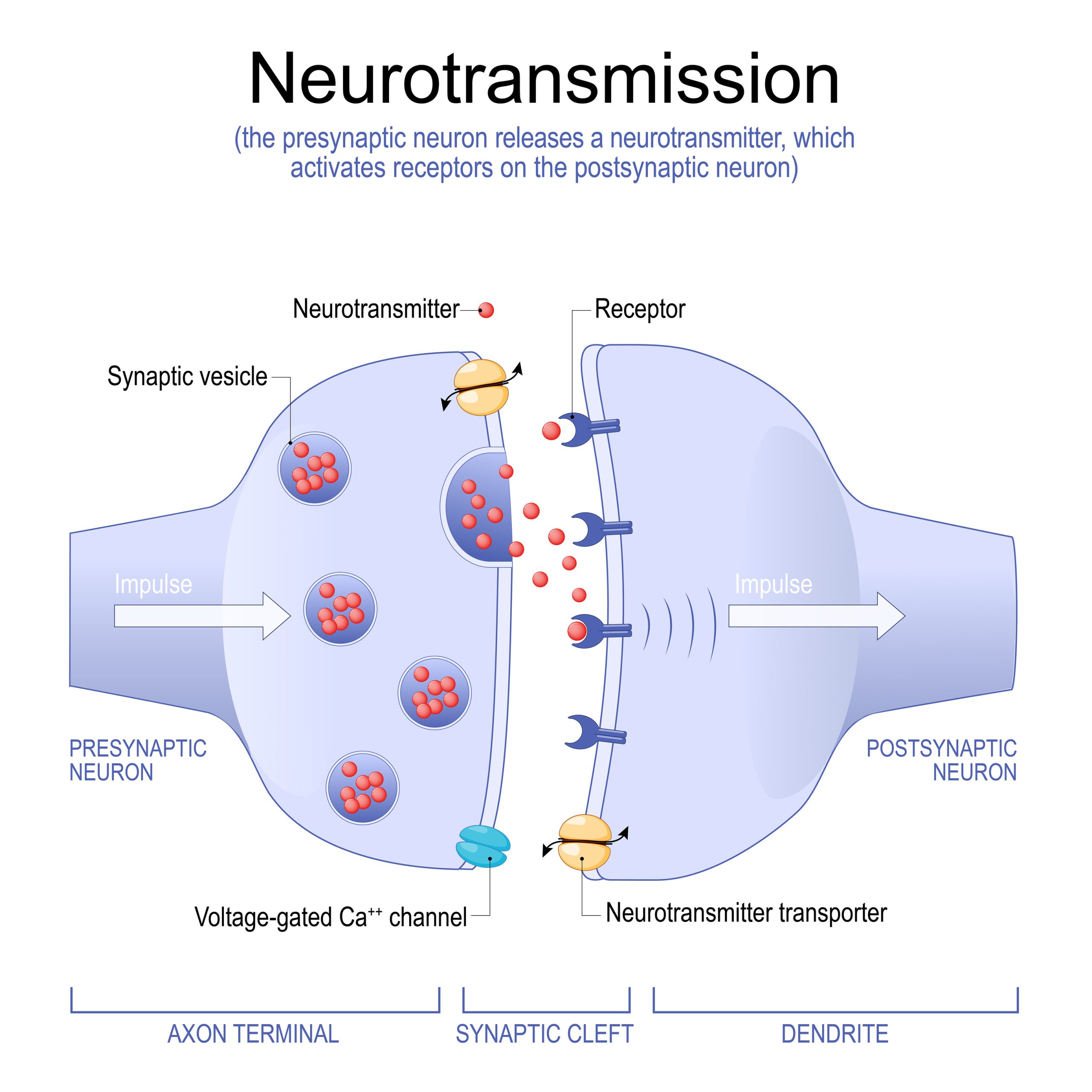
Neurotransmission is the process by which neurons communicate with each other, allowing for the transmission of signals throughout the nervous system. This complex process is fundamental to all neuronal functions, from the basic motor responses to sophisticated cognitive processes. Neurotransmission occurs at synapses, specialized junctions where neurons come into close proximity but do not physically touch. The synaptic cleft, a small gap between the presynaptic neuron (sending neuron) and the postsynaptic neuron (receiving neuron), is where the transfer of information occurs through chemical messengers known as neurotransmitters (Kandel, Schwartz, Jessell, Siegelbaum, & Hudspeth, 2013).
The process begins when an action potential, or an electrical signal, reaches the synaptic terminals of the presynaptic neuron. This electrical signal triggers the influx of calcium ions through voltage-gated calcium channels. The increased concentration of calcium inside the neuron prompts vesicles filled with neurotransmitters to fuse with the presynaptic membrane and release their contents into the synaptic cleft. Neurotransmitters then diffuse across the cleft and bind to specific receptors located on the membrane of the postsynaptic neuron. The binding of neurotransmitters to these receptors can activate them, causing either an excitatory or inhibitory effect on the postsynaptic neuron, depending on the nature of the neurotransmitter and the receptor (Bear, Connors, & Paradiso, 2020).
Excitatory neurotransmitters, such as glutamate, typically cause the postsynaptic neuron to become more likely to fire an action potential by depolarizing the neuron, reducing the membrane potential. Conversely, inhibitory neurotransmitters, like GABA, increase the membrane potential, making the postsynaptic neuron less likely to fire an action potential. This balance between excitation and inhibition is critical for the healthy functioning of the brain and the prevention of neurological disorders. Additionally, neurotransmitters can be degraded by enzymes in the synaptic cleft or taken back into the presynaptic neuron through reuptake channels, ensuring that the signal is brief and precisely controlled (Purves et al., 2018).
Understanding the dynamics of neurotransmission is crucial for comprehending how the brain processes information, regulates bodily functions, and interacts with the environment. Disruptions in neurotransmission can lead to various neurological diseases, such as depression, schizophrenia, and Parkinson’s disease, highlighting the importance of neurotransmitters in maintaining neural and overall health.
Neurochemical Regulation
Neurochemical regulation, particularly the role of neurotransmitters, is foundational to understanding resilience in individuals. Neurotransmitters, being the chemical messengers of the nervous system, regulate various brain functions and, by extension, influence behaviors, moods, and cognitive processes (Russo et al., 2012). Among these neurotransmitters, serotonin, dopamine, and norepinephrine have shown significant implications for resilience. Additionally, the brain’s endogenous opioid system, involved in pain and pleasure, has been associated with coping and resilience (McEwen, 2007).
Serotonin, often referred to as the “feel-good” neurotransmitter, plays a pivotal role in mood regulation (Hensler, 2006). Serotonin influences a myriad of psychological processes including mood, anxiety, and aggression. A balance in serotonin levels is crucial for mental health and well-being, while an imbalance might contribute to mood disorders such as depression. Additionally, variations in genes related to serotonin production, reuptake, and reception might contribute to individual differences in resilience. Specifically, certain polymorphisms in the serotonin transporter gene (5-HTT) have been linked to greater vulnerability to stress and lower resilience (Caspi et al., 2003). Such genetic nuances underscore the profound connection between serotonin pathways and resilience, suggesting that certain individuals, due to their genetic makeup, might naturally possess or lack resilience mechanisms against psychological adversities.
Dopamine, another key neurotransmitter, is predominantly associated with the brain’s reward system. Dopaminergic pathways play a vital role in motivation, pleasure, and reward-seeking behaviors (Wise, 2004). Resilience often requires individuals to engage in goal-directed behaviors despite setbacks and challenges. A well-functioning dopamine system ensures motivation is maintained during these adversities. Reduced dopaminergic activity, on the other hand, can lead to anhedonia (lack of pleasure) and diminished motivation, thereby hampering resilience. It’s also relevant to recognize the balance of dopamine production; excessive amounts can result in impulsive behaviors or even addiction, underscoring the importance of balanced neurochemistry for adaptive resilience.
Norepinephrine (or noradrenaline), primarily involved in the fight-or-flight response, has implications for alertness, attention, and stress response. Elevated levels of norepinephrine during stressful events can sharpen attention and focus, facilitating better decision-making and adaptive responses (Aston-Jones & Cohen, 2005). However, chronically elevated levels, as seen in prolonged stress, might impair resilience, leading to burnout and cognitive fatigue.
Beyond these primary neurotransmitters, the endogenous opioid system of the brain, which regulates pain and pleasure, also plays a role in resilience. Opioids, by mediating feelings of pain and pleasure, influence coping mechanisms. A balanced endogenous opioid system ensures effective pain modulation and emotional coping during adversity, adding another layer of complexity to the neurochemical landscape of resilience (McEwen, 2007).
Understanding the neurochemical basis of resilience offers a deeper insight into why some individuals bounce back from adversities with ease, while others struggle. This knowledge opens avenues for targeted interventions, which can help enhance resilience by modulating neurotransmitter pathways.
Neurobiological Mechanisms Involved in Resilience
One of the most compelling areas of research in the realm of resilience is the exploration into the neural mechanisms that facilitate adaptive responses to adversity. Over the past few decades, groundbreaking work has shed light on how our brain and its intricate networks enable resilience, allowing individuals to overcome, adapt to, and grow from challenges (Southwick & Charney, 2012).
Neural Plasticity and Neurogenesis
At the heart of resilience lies the brain’s plastic nature, its ability to change and adapt in response to experiences. Neural plasticity is not just about brain changes during the early developmental years but extends across the lifespan (Merzenich, 2013). Adaptive plasticity permits the brain to reorganize and strengthen neural pathways, thus equipping individuals to cope better with future adversities. Neurogenesis, particularly in the hippocampus, plays a role in cognitive flexibility and emotional responses, thereby influencing resilience (Kempermann, 2019).
Adaptive Plasticity and Its Role in Resilience
Adaptive neural plasticity refers to the brain’s remarkable ability to restructure and modify its own neural pathways based on experiences. This dynamic attribute of the brain is not just a reaction to learning or environmental changes, but a proactive process that equips an individual to handle novel situations and challenges more effectively (Holtmaat & Svoboda, 2009). When the brain encounters new information or experiences traumatic events, it undergoes structural and functional alterations. This includes the strengthening of certain synaptic connections, while others may weaken or dissipate entirely. Such changes in synaptic strength, known as synaptic plasticity, are believed to be the foundation for learning and memory (Citri & Malenka, 2008).
In the context of adversities, adaptive plasticity acts as a protective mechanism. When faced with stressful situations or traumas, the brain’s ability to reorganize itself allows individuals to process, adapt to, and eventually overcome these experiences. For instance, following a traumatic event, certain neural pathways associated with the trauma might become overly active, leading to heightened stress responses. However, with the right interventions or coping strategies, the brain can reorganize these pathways to reduce the intensity of these responses and promote a return to a more balanced state (McEwen, Nasca, & Gray, 2016).
Additionally, adaptive plasticity enhances an individual’s ability to learn from past experiences. This learning doesn’t merely involve factual recall or procedural habits, but extends to emotional learning and behavioral adaptability. By strengthening neural pathways that led to successful coping in the past, the brain better prepares itself for similar future adversities. In this way, adaptive plasticity is intrinsically linked to resilience. It provides a neural basis for why some individuals can bounce back from adversities while others might struggle more significantly (Davidson & McEwen, 2012).
The brain’s adaptive plastic nature is central to human adaptability and resilience. By continuously reorganizing and strengthening neural pathways based on past experiences, it equips individuals with the tools necessary to cope more effectively with future challenges and adversities.
Neurogenesis and Its Influence on Cognitive Flexibility and Emotional Responses
Neurogenesis, the process of forming new neurons, is a dynamic occurrence predominantly observed in the hippocampus, a region of the brain linked to various cognitive and emotional functions. One of the significant outcomes of neurogenesis is its influence on cognitive flexibility, which is the brain’s ability to adapt to new information and shift between different tasks or thoughts (Kempermann, 2008). As new neurons integrate into existing neural networks within the hippocampus, they foster a neural environment that promotes adaptability and learning. For example, newly formed neurons contribute to pattern separation, a mechanism by which similar experiences or memories are kept distinct (Sahay et al., 2011). This capability is crucial for an individual to adapt to changing environments and situations, facilitating improved decision-making processes and problem-solving abilities.
Emotionally, the hippocampus has a significant role in regulating responses, particularly in the context of stress and trauma. The newly generated neurons in the hippocampus have been found to be especially sensitive to experiential factors, suggesting that they play a vital role in modulating emotional responses (Anacker & Hen, 2017). When faced with stressors, an increase in hippocampal neurogenesis can aid in the attenuation of stress responses, promoting a more balanced and measured emotional reaction. This could, in part, explain why some individuals recover more rapidly from emotional setbacks or traumas, as a more robust rate of neurogenesis may provide a buffer against the deleterious effects of stress, thereby promoting resilience (Schoenfeld & Gould, 2012).
In essence, the role of neurogenesis, particularly in the hippocampus, cannot be understated when it comes to cognitive flexibility and emotional regulation. By facilitating a dynamic and adaptable neural environment, neurogenesis allows for improved cognitive adaptability and more balanced emotional reactions, key components in the foundation of resilience. A healthy rate of neurogenesis is, therefore, an essential factor in enhancing an individual’s ability to cope with and recover from (both cognitively and emotionally) adversities, particularly those that constitute novel situations.
Time Out for Reflections on Resilience . . .
How can our understanding of the neurobiology of resilience be translated into real-world applications, such as therapy, education, or workplace training?
Stress, the Brain, and Resilience: An Overview
Resilience, broadly defined, represents an individual’s ability to withstand and bounce back from adversity, and in certain circumstances, to grow and thrive in the face of challenges (Masten, 2001). This concept, deeply embedded in the tapestry of human survival and evolution, has been researched extensively in the context of psychology. However, contemporary studies also direct our attention to the intricate workings of the brain when investigating resilience. The synergy between stress, the brain’s response mechanisms, and the manifestation of resilience is a cornerstone in understanding the neurobiology of resilience.
Resilience is not merely a passive shield against stress; it’s an active process, a dynamic interplay of molecular, cellular, and neural circuitry components that synergistically act to maintain equilibrium and mental health (Feder et al., 2009). As stress cascades through the brain’s neural networks, resilience counterbalances, protecting and enabling the individual to navigate through the stormy seas of psychological distress and environmental challenges.
The brain, as the central organ mediating our perception and response to environmental stressors, employs complex neural pathways and signaling mechanisms in response to stress. The initiation of the stress response begins with the amygdala, which identifies potential threats and subsequently activates the hypothalamus. The hypothalamus, in turn, triggers the hypothalamus-pituitary-adrenal (HPA) axis, resulting in the release of stress hormones, notably cortisol (Herman et al., 2005). While the acute release of cortisol prepares the body to confront or escape the threat (commonly known as the “fight or flight” response), chronic activation of this system, due to prolonged exposure to stress, can lead to deleterious effects on multiple body systems, including the brain (McEwen, 2007).
Yet, not all individuals exposed to stressors develop pathological symptoms or suffer from chronic stress-related disorders. This variation can be attributed to resilience, a complex interplay of genetic, epigenetic, and environmental factors (Russo et al., 2012). At the core of neurobiological resilience is the brain’s ability to engage in neural plasticity, allowing adaptive changes in neural pathways and synapses in response to experiences (Davidson & McEwen, 2012). Additionally, certain brain regions, such as the prefrontal cortex, modulate the stress response by exerting inhibitory control over the amygdala, regulating emotional responses and facilitating adaptive behavior (Arnsten, 2009).
A deeper dive into this subject reveals that the brain’s resilience mechanisms are not just about suppressing negative responses. The release of growth factors like brain-derived neurotrophic factor (BDNF) in response to certain stressors can promote neural growth and connectivity, underscoring the brain’s ability to adapt and even thrive amidst challenges (Duman & Monteggia, 2006).
Resilience is underpinned by a myriad of neurobiological processes that are simultaneously intricate and adaptive. Understanding these processes lays the groundwork for therapeutic interventions and strategies to foster resilience, especially in populations at risk of stress-related disorders.
Stress Response Systems and Resilience
The human body’s stress response systems have evolved as protective mechanisms to help us respond to environmental challenges and potential threats. At the heart of this system is the hypothalamic-pituitary-adrenal (HPA) axis. Under normal circumstances, this axis operates in a tightly regulated feedback loop. In the presence of a perceived threat or stressor, the hypothalamus releases corticotropin-releasing hormone (CRH), which in turn stimulates the anterior pituitary gland to secrete adrenocorticotropic hormone (ACTH). ACTH then prompts the adrenal cortex to produce and release cortisol, a glucocorticoid hormone that has wide-ranging effects on various bodily functions, from modulating the immune response to influencing metabolic processes and cognition (McEwen, 2007).
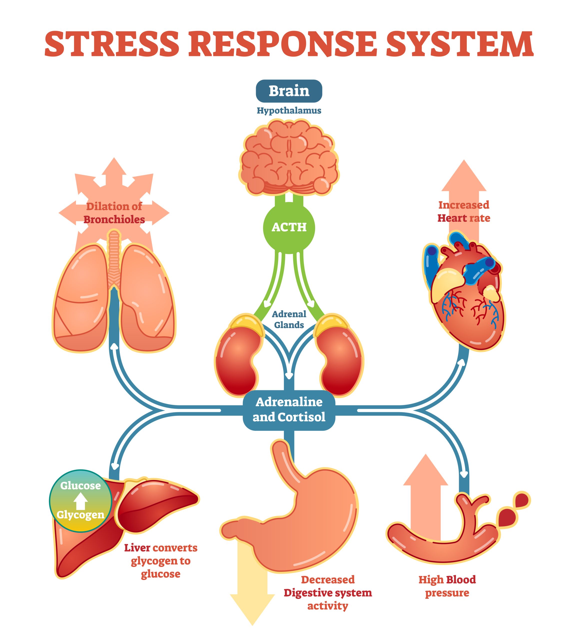
Acute activation of the HPA axis, and the subsequent release of cortisol, prepares the body to respond effectively to short-term challenges. This so-called “fight or flight” response enhances alertness, provides a burst of energy, and temporarily suppresses non-essential functions, such as digestion and reproduction. In this way, cortisol plays a pivotal role in allowing the organism to respond adaptively to its environment (McEwen & Gianaros, 2011). However, while acute stress reactions can be adaptive, problems arise when the HPA axis is chronically activated or dysregulated. Continuous exposure to high levels of cortisol can have deleterious effects on almost every system in the body. Neurologically, chronic stress can result in atrophy in areas of the brain vital for memory and cognitive functions, particularly the hippocampus. This neurobiological damage can pave the way for the onset of various psychiatric disorders, including depression, anxiety disorders, and post-traumatic stress disorder (Sapolsky, 2004).
Nevertheless, it’s crucial to recognize that not everyone exposed to chronic stress or trauma develops psychiatric disorders. As noted, resilience – this ability to bounce back from adversity and maintain functional psychological and physical health – varies among individuals (Southwick, Bonanno, Masten, Panter-Brick, & Yehuda, 2014). Factors contributing to resilience are manifold and interwoven. Adaptive regulation of the HPA axis is one key biological underpinning. However, socio-psychological factors play an equally, if not sometimes more, significant role. The presence of robust social support networks has been consistently identified as a protective factor against the detrimental effects of stress. Such networks provide emotional sustenance, as well as cognitive resources, that can mitigate the negative impacts of adversity (Charney, 2004). Additionally, learned coping mechanisms, whether through personal experiences or interventions, can equip individuals with the tools to navigate challenges more effectively. Such skills might include cognitive restructuring, problem-solving techniques, and emotion regulation strategies (Masten & Narayan, 2012).
While the neurobiological underpinnings of resilience are intricate and multifaceted, understanding them offers valuable insights into how some individuals weather storms with grace and tenacity. It provides a roadmap for interventions aimed at fostering resilience at both clinical and community levels.
Time Out for Reflections on Resilience . . .
Can you think of a time when you displayed resilience in the face of adversity?
Based on the reading, what neurobiological processes might have been at play?
Case Studies: Stress and Resilience
The exploration of stress and resilience through case studies provides a concrete, humanized understanding of the theoretical and neurobiological concepts discussed earlier. These case studies, often grounded in real-life scenarios, offer insights into the complex interplay of biological, psychological, and environmental factors in resilience.
Case Study 1: Post-Traumatic Growth in Military Veterans
One profound example comes from studies involving military veterans, a group often exposed to extreme stress and trauma. A study by Feder et al. (2013) examined veterans who had experienced combat-related trauma. Remarkably, a subset of these individuals demonstrated significant post-traumatic growth, an increased sense of personal strength, and improved interpersonal relationships post-deployment. Neuroimaging and hormonal assessments revealed that these veterans exhibited unique patterns in their neural circuitry, particularly in regions associated with emotional regulation and cognitive control. These findings suggest that exposure to severe stress does not inevitably lead to dysfunction; instead, it can catalyze significant personal growth and heightened resilience.
Case Study 2: Resilience in Children with Adverse Childhood Experiences (ACEs)
Another compelling case study involves children with high ACE scores who demonstrate remarkable resilience. A longitudinal study by Bethell et al. (2014) tracked children with multiple ACEs, observing how certain factors, like supportive adult relationships, contributed to resilience. Neurobiological assessments revealed that these resilient children showed adaptive changes in their stress response systems, including more regulated cortisol levels and enhanced neural plasticity. This case study underscores the potential for positive environmental factors to mitigate the adverse neurobiological impacts of early life stress.
Case Study 3: The Role of Community and Culture in Resilience among Indigenous Populations
Research on indigenous populations, such as the work of Kirmayer et al. (2011), offers valuable insights into the collective and cultural aspects of resilience. Despite facing significant socio-economic and health disparities, many individuals in these communities exhibit a strong sense of resilience. This resilience is often rooted in cultural practices, community support, and a deep connection to heritage and land. Neurobiological studies in these populations have highlighted the role of social networks and cultural engagement in modulating stress response systems, thereby fostering resilience.
These case studies emphasize the multifactorial nature of resilience. They illustrate that resilience is not solely an individual trait but is deeply influenced by interpersonal relationships, community support, and cultural factors. Furthermore, they highlight the potential for neurobiological mechanisms to adapt positively in the face of adversity, offering hope for interventions aimed at enhancing resilience in various populations.
Media Attributions
- vintage-1418613_1280 © ArtsyBee
- anatomy-function-brain-areas-basics-aug-2019-2024
- Cross section through the brain showing the limbic system and all related structures
- Diagram of Neuron Anatomy © colematt
- Synapse Structure. Neurotransmitter, synaptic vesicles and synaptic cleft. © ttsz
- Stress response system vector illustration diagram, nerve impulses scheme.
The process and outcome of successfully adapting to challenging or threatening experiences, especially through mental, emotional, and physical resistance or elasticity.
A state of mental or emotional strain resulting from challenging or adverse situations. Physiologically, stress activates the body's "fight or flight" response, releasing hormones like cortisol and adrenaline.
A network of brain structures crucial for regulating emotions, forming memories, and governing behaviors.
A part of the brain involved in processing emotions, particularly those related to fear. It plays a significant role in triggering the body's response to stressful or threatening situations.
A region of the brain associated with learning, memory, and emotion regulation. Chronic stress can negatively impact the hippocampus, potentially leading to memory problems and mood disorders.
How neurotransmitters, the chemical messengers of the nervous system, influence resilience by affecting behaviors, moods, and cognitive processes.
Chemical messengers that transmit signals across synapses from one neuron to another. They play a crucial role in determining mood, emotions, and other psychological processes.
A neurotransmitter involved in mood, anxiety, and aggression regulation. Imbalances can affect mental health and resilience.
A neurotransmitter associated with motivation, pleasure, and reward-seeking behaviors.
A neurotransmitter involved in alertness, attention, and stress response.
Brain system regulating pain and pleasure, influencing coping mechanisms during adversity.
Techniques or strategies that individuals use to manage and adapt to challenging situations.
The proactive ability of the brain to modify its neural pathways based on experiences, thereby preparing an individual to handle novel situations and challenges effectively.
The formation of new neurons, primarily in the hippocampus, influencing cognitive flexibility and emotional responses.
Changes in the strength of synapses, believed to be the basis for learning and memory.
The brain's ability to switch between different tasks or thoughts and adapt to new information.
A mechanism allowing the brain to distinguish between similar experiences or memories.
The study of the nervous system's cellular and molecular biology, especially concerning the brain and its influence on behavior and cognitive functions.
A steroid hormone produced by the adrenal glands, often in response to stress. While it has many functions, in the context of stress, cortisol helps the body respond effectively, but chronic elevation can be detrimental.
A physiological reaction in response to perceived harmful events, which prepares the body to either confront or flee from the threat.
The ability of the brain to reorganize itself by forming new neural connections. This adaptability is essential for learning, memory, and recovery from injury.
A junction between two nerve cells where neurotransmitters are released to allow signals to pass from neurons to other cells.
The front part of the frontal lobe, this brain region is involved in executive functions such as decision-making, planning, and moderating social behavior. It also plays a role in modulating emotional responses from the amygdala.
A protein in the brain that supports the survival, growth, and differentiation of neurons. BDNF is crucial for long-term memory and plays a role in neural plasticity, which is the brain's ability to adapt and change over time.
A complex set of interactions between the hypothalamus, the pituitary gland, and the adrenal glands. This system plays a key role in the body's response to stress.
Interpersonal relationships that provide emotional and cognitive resources, identified as protective factors against the effects of stress.

