8 Measuring toxicity of Lunar and Martian regolith simulants on bacteria
Hugo Castillo and Makaila Olson
Measuring toxicity of Lunar and Martian regolith simulants on bacteria
Preparation of the overnight culture (Done by your TA)
- Add 30 mL of nutrient broth (NB) to a 50 mL conical tube.
- From an NB reference agar plate of Escherichia coli, use an inoculating loop to pick up a single colony.
- Place the loop in the 50 mL conical tube of broth and stir it to release the cells.
- Discard the inoculating loop as biohazard waste.
- Close the tube and seal with a strip of parafilm.
- Place the 50 mL conical tube on a rack inside a microbiological incubator set at 30°C.
- Incubate the cells for 7 hours.
Preparation of the simulant (Adapted tyndallization protocol) (Done by your TA)
- Place 20 g of lunar simulant (e.g. LHS-1) in an Erlenmeyer flask. Cover the top with foil.
- Place the flask in the autoclave and run a regular cycle (15 min at 121°C). Once the cycle is complete, take the flask out of the autoclave and allow the materials to cool to room temperature before proceeding.
- Transfer the flask to a microbiological incubator set to 37°C and incubate for 24 hours.
- After incubation, place the flask back into the autoclave and repeat step 2.
- Allow the materials to cool to room temperature before proceeding.
- Place 40 2 mL Eppendorf tubes and the sterile flask in a biosafety cabinet.
- While in the biosafety cabinet, transfer approximately 0.5 g of LHS-1 into each tube and close them immediately.
- Once the simulant is fully aliquoted, transfer the tubes to the autoclave and repeat step 2, except for the autoclave inc
- Allow the materials to cool to room temperature before commencing experimental procedures.
Overnight culture concentration and preparation
- Label two 2-mL Eppendorf tubes as treatment and control.
- Transfer 1.9 mL of the overnight culture into each tube.
- Centrifuge at 10,000 RPM for 5 minutes to pellet the cells.
- Carefully remove the supernatant without disturbing the pellet.
- Wash the cells by adding 1.5 mL of 0.9% saline to each tube.
- Vortex thoroughly to resuspend the cell pellets until no visible clumps remain.
- Centrifuge the tubes at 10,000 RPM for 5 minutes.
- Carefully remove the supernatant from each tube.
- Add another 1.5 mL of 0.9% saline to each tube. Do not vortex during this step. Gently mix the saline with the pellet by inversion if necessary.
- Centrifuge the tubes at 10,000 RPM for 5 minutes.
- Remove the supernatant carefully to ensure the pellets remain intact.
- Resuspend the pellets in 1 mL of 0.9% saline per tube.
- Vortex thoroughly to ensure the cells are evenly resuspended.
Simulant exposure
- Transfer the resuspended cells into a tube with regolith simulant (treatment) or leave them in the tube (control).
- Invert the simulant tubes several times to ensure proper mixing of the substrate and the cells.
- Attach the treatment tubes flat against the front-facing wheel of a clinostat or in another type of cell rotator or mixer. Put the control tubes on the gravity axis of the clinostat.
- Place the clinostat inside a microbiological incubator set at 30°C.
- Turn on the clinostat and ensure the speed is set to 8 rpm. Incubate for 48 h.
- At the end of the incubation, remove the tubes from the clinostat and process immediately.
Cell count measurement (colony-forming units per mL)
This process involves a serial dilution up to 10-8, therefore 8 sterile Eppendorf tubes will be needed per sample.
- Calculate the number of Eppendorf tubes needed by multiplying the number of samples that will be used for CFU measurement by 8. For example, if you are working with six samples, you will need 48 Eppendorf tubes.
- Fill each Eppendorf tube with 900µL of 0.9% saline solution.
- For each tube set, label each tube 1 to 8.
- Starting with the first sample, add 100 µL of the cell culture to the tube # 1.
- Vortex the tube at maximum speed for 5 seconds, then transfer 100µL of the mixture into tube # 2.
- Repeat step 5 until the last tube.
- Using a permanent marker, divide the back of a NB agar plate into four sectors and label as follows:
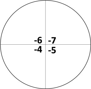
- Starting with the tube # 4, transfer 10 µL aliquots of the cell’s dilution onto the corresponding quadrant. Your instructor will provide specific directions on the dilutions needed for your experiment.
- Repeat twice more for a total of three drops in the same quadrant.
- Repeat steps 8 and 9 with tubes # 5, # 6, and # 7.
- Let the plate sit at room temperature until the liquid has completely absorbed into the agar. Handle the plates with care to prevent the liquid drops from mixing and/or spreading on the plate.
- Incubate the plates at 30°C for a minimum of 12 hours, but not more than 15. If the plates are incubated for too long, the colonies will overgrow.
- Repeat steps 4-12 with the remaining samples.
- After incubation, identify the quadrant that has a countable number of colonies, this is, that where each drop grew between 10 and 30 individual colonies. If none of the quadrants have enough colonies, incubate the plates 2 to 4 more hours. If all of the quadrants have to many colonies to count, the plate was left in for too long.
- Once a quadrant has been selected, count and record the number of colonies in each drop. Calculate the average number of colonies on the quadrant and use the following equation to calculate CFU:
where the # of colonies is the average that was calculated, the volume of the drop is the amount of mixture added from the tube onto the plate (10µL or 0.01 mL), and the quadrant dilution is 10 to the power of the quadrant number (quadrant -5 would be ). The value calculated corresponds to the number of colony- forming units per mL.
- Repeat steps 14 to 16 for the remaining plates.
Biofilm biomass (crystal violet assay)
- Remove the planktonic cells (free swimming cells) from the tubes using a micropipetor set at 1 mL. Make sure you remove as much buffer as possible.
- Carefully add 1 mL of 0.9% saline solution to each tube. Do not mix the tube.
- Remove the saline as described on step 1 and repeat the rinsing procedure for a total of three times.
- Carefully add 1 mL of 0.1% crystal violet stain to each tube and let sit for 5 minutes.
- After 5 minutes, remove the crystal violet from all tubes following the procedure previously described. Make sure to deposit the crystal violet into a container designated for chemical waste.
- Carefully rinse the tube with 1-mL of 0.9% saline solution for a total of 5 times. Mix the stimulant with the buffer on each wash.
- Following the washes, 1- mL of 91% isopropyl alcohol to the tube to dissolve the stained biofilms.
- Once dissolved, transfer 100 µL aliquots, in triplicate, into separate wells of a 96-wells plate and measure absorbance 550 nm using a spectrophotometer or a plate reader. The absorbance value is directly related to the biomass of the biofilm developed in the tubes.